Earlier translational safety
insights through
human-relevant
multi-tissue systems
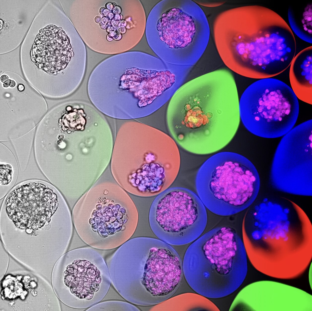
Efficacy, bioactivation and organ-specific toxicity, assessed together in one human assay. Tumour, liver and cardiac tissue share a well, systemic drug exposure is emulated, and more than 1,500 proteins are read out per sample. Every safety signal arrives with its mechanism attached.
Find the liability while you can still design around it
Toxicity risks are still detected too late. Traditional in vitro systems often lack systemic relevance, and single-tissue assays miss the three things that decide a programme: how organs influence each other, how a compound is activated or inactivated by the liver, and how tissues respond to stress before any cells die.
Organ liabilities surface late
Cardiac and hepatic effects often appear in vivo or in the clinic, once considerable budget and time have been spent.
Single-tissue models miss
system interactions
A tumour cell line alone will not bioactivate a prodrug. Without liver tissue in the well, you do not measure the true potency of the molecule.
Viability alone does not tell you
the mechanism
A cell death curve shows efficacy or toxicity. In-depth protein profiling shows which pathways became active or inactive, in which tissue, at which dose.
Two platforms, one experiment
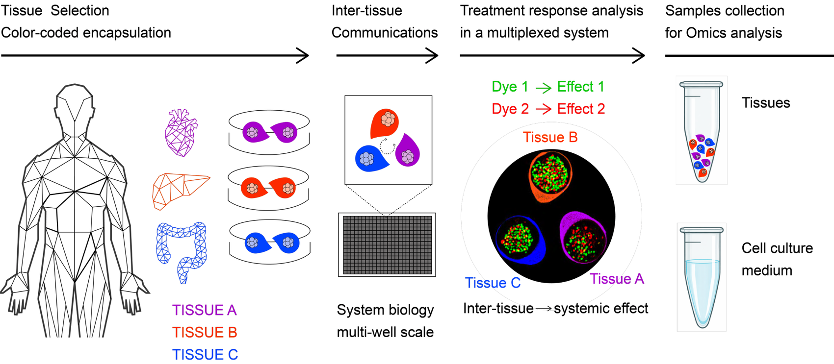
A human 3D multi-tissue culture supplies the biology; affinity proteomics supplies the depth. Encapsulated tumour, liver and cardiac tissues are co-cultured in one well, dosed together, and read out for cell death and for more than 1,500 proteins from the same experiment.
FluoXplore: several human tissues, one well, one exposure
Colour-coded encapsulation keeps each tissue identifiable while they share medium, so inter-tissue communication and bioactivation happen inside the assay instead of being inferred from separate experiments. Treatment response is read per tissue in a multiplexed system at multi-well scale.
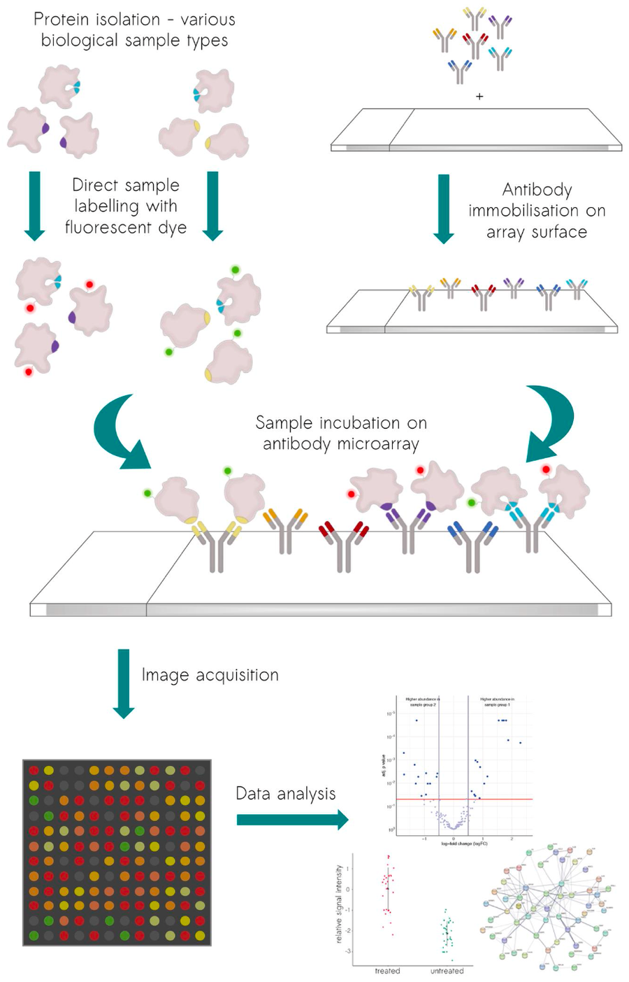
scioDiscover®: more than 1,500 proteins from the same sample
Proteins from supernatants and cell pellets are labelled directly and incubated on antibody microarrays. Because every protein is measured in every sample, dose series stay comparable and no values have to be imputed. What arrives is not a list of hits but a signature that maps to pathways.
Tissue generation and encapsulation
Tissue combination chosen for the question, encapsulated and co-cultured.
Multi-tissue dosing and imaging
One exposure across all tissues, with phenotypic response read per tissue.
Supernatant and pellet proteomics
Secreted and intracellular proteins from the same wells, profiled in parallel.
Integrated analysis and interpretation
Phenotype and proteome read together, not in two separate reports.
The multi-tissue biology is delivered with FluoXplore on FluoSphera's Pleiad platform; protein profiling, statistics and biological interpretation are delivered by Sciomics, based on the scioDiscover® platform.
What a broad protein readout adds
A viability curve tells you the potency, efficacy or toxicity. A proteome-wide readout tells you which mechanism was involved, in which tissue, and how far below the toxic dose it started.
Proteins specific to the mechanism of action of the tested compound move first, showing whether the compound reaches its target and acts on it.
Acute-phase and hepatocyte stress markers point to genuine hepatotoxicity rather than a transient metabolic burden.
Cardiac stress and remodelling proteins give an early, sub-lethal warning where late failures are most expensive.
Signatures, not single analytes, are what survive contact with a real programme. The colour code follows the tissues throughout this page.
A study designed around your decision, not around our catalogue
Study design
Tissue combination, dose range and sampling points chosen for your question, with a dedicated scientist from the first call on both sides.
Small samples, full depth
Supernatants and cell pellets from standard plate formats. Four technical replicates, average CV under 10 %, no missing values across the data matrix.
Decision-ready report
Statistics, pathway mapping and biological interpretation, publication-ready and structured for a go/no-go rather than a data dump.
Timelines: three to six weeks for the proteomic part. The total duration depends on model complexity and treatment schedule. What you get is the pace of two committed specialists rather than a queue inside a large organisation.
A prodrug that needs the liver
Capecitabine only becomes active once the liver has converted it to 5-fluorouracil, and it carries known cardiac and hepatic liabilities. That makes it a concrete test case for whether a human multi-tissue assay behaves the way a patient does.
The integrated workflow was applied to this compound as a proof of concept and presented together at Swiss Biotech Day 2026, in a study run with a global pharmaceutical company. Bioactivation, the resulting therapeutic window and the mechanistic biomarker panels are described in the poster.
The full poster, including all figures, is available on request.
Relevant for complex preclinical development challenges
Oncology
Complex efficacy and toxicity relationships.
ADCs
Payload-driven and off-target toxicity.
Small molecules
Liabilities that only appear after hepatic bioactivation.
Immunology
Systemic and tissue-specific responses.
Combination therapies
Organ interaction and integrated response profiling.
Biologics
Secreted response profiles alongside functional readouts.
Go deeper on either side
of the workflow
Human multi-tissue systems
Affinity proteomics for preclinical research
Two independent companies,
one combined service offering
FluoSphera contributes human multi-tissue systems and systemic phenotypic profiling. Sciomics contributes high-content proteomic profiling, statistics and biomarker interpretation. Studies are scoped jointly, and you talk to the scientists who run them.
 FluoSphera SA · Geneva, Switzerland
 Sciomics GmbH · Neckargemünd, Germany
Tell us what you need to decide
We will tell you honestly whether a human multi-tissue study with a broad proteomic readout can answer it, and what it would take.
Both buttons open an email to a shared inbox read by both companies. No form, no tracking, no newsletter sign-up.